Neuro-MS.NET

Présentation du Neuro-MS.NET
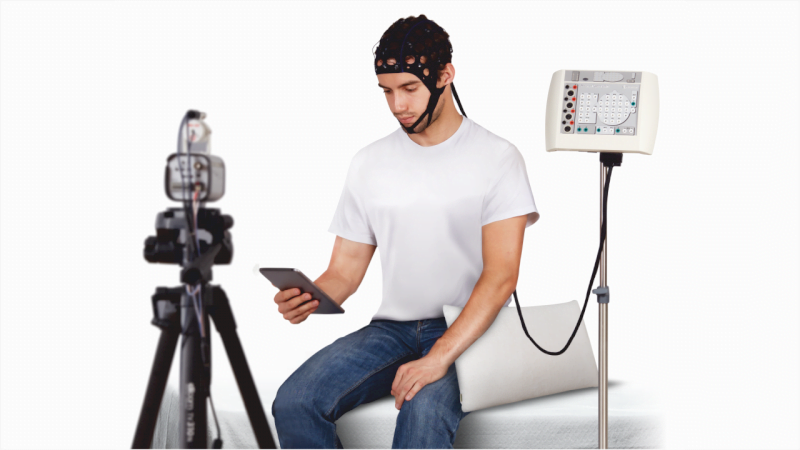
The stimulator can be controlled by a Windows computer equipped with Neuro-MS.NET software. The interface between the computer and the central unit is via a simple USB port.
The Neuro-MS.NET software includes the “patient” databases, a library and editor of treatment protocols, and the TMS machine control system.
The software guides the user through regular tutorials explaining, for example, how to register a new patient, select a predefined protocol from the library, create or edit a new treatment protocol, perform or terminate a stimulation session, display the detailed history of each treatment on the screen and print a treatment report.
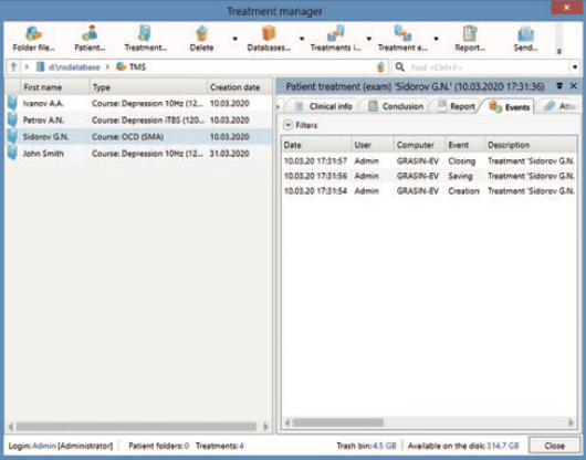
Patient database
The “patient” database contains a list of all patients and the history of all treatments.
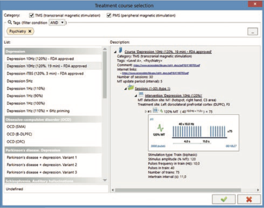
Predefined protocols
The software offers a large number of predefined treatment/rehabilitation protocols. The user can create new protocols or modify any parameter of the existing ones at any time.
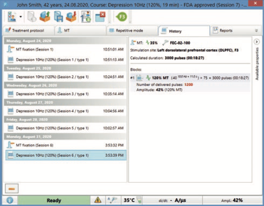
History
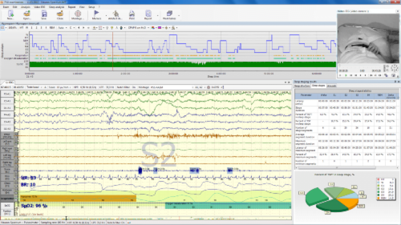
The treatment history stores the data obtained during the determination of the motor threshold (including potentials), the data of the treatment sessions performed, the date of the session start, the actual number of stimuli applied in each session and many other data.
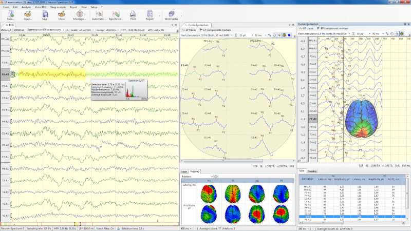
Motor threshold (MT) determination and brain mapping tools
The measurement of the motor threshold (MT) makes it possible to determine the “dosage” of the rTMS treatment. This is an important measure for most rTMS protocols. Accuracy of TM measurement is the key to achieving treatment efficacy and safety. As well as locating the treatment site, the determination of the TM must be done quickly but accurately. Neuro-MS.NET software offers a set of tools for both MT determination and brain mapping: automatic MT determination using an EMG amplifier, semi-automatic MT determination using STEP algorithms, the F3 marker and visual aid.
Automatic determination of the motor threshold
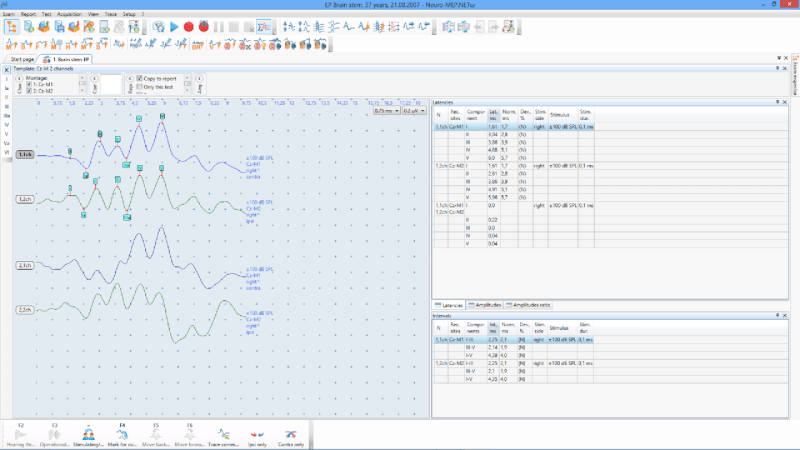
The motor threshold can be determined automatically using a compatible EMG amplifier. In this mode, the software automatically sends a series of pulses at random intervals. A high-performance algorithm, specially designed for this purpose, gradually increases or decreases the intensity by following the amplitude of the specific EMG response. In a few steps, the algorithm automatically finds the threshold value.
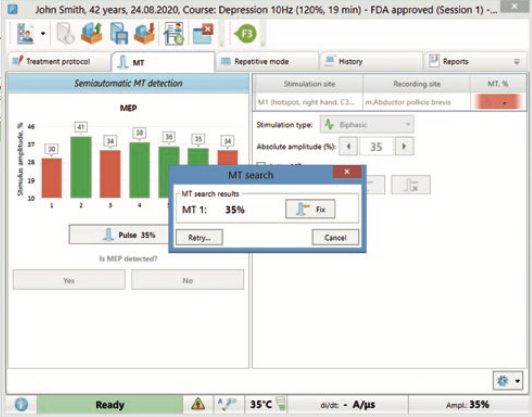
Semi-automatic determination of the motor threshold
In semi-automatic mode, the software also sends a series of impulses, increases or decreases the intensity of the stimulus according to the respective responses given by the user who visually observes the contraction of the muscle and indicates this response to the software by clicking “yes” or “no”. The motor threshold is usually determined in only 6 to 8 steps per stimulus. This approach simplifies the determination of the motor threshold and guarantees high accuracy and speed.
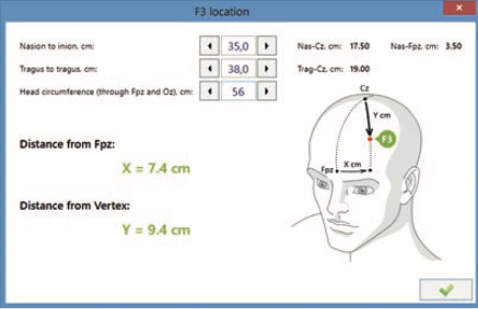
Once the motor threshold is determined, it is important to position the coil correctly over the stimulated area. The conventional protocol for the treatment of depression involves stimulation of the left dorsolateral prefrontal cortex (DLPFC), which corresponds to the F3 point in the 10-20 system.
The manual location of the F3 requires a lot of measurements and calculations. Our software is equipped with an algorithm for F3 localization that uses only 3 measurements:
– The distance between tragus,
– The distance between the nasion and the inion,
– The circumference of the head.
Simply enter these measurements and the software calculates the exact target point. More precise mapping of the brain can be obtained with the MRI-guided neuro-navigation system that can be installed on Neuro-MSX.
Marker F3
Once the motor threshold is determined, it is important to position the coil correctly over the stimulated area. The conventional protocol for the treatment of depression involves stimulation of the left dorsolateral prefrontal cortex (DLPFC), which corresponds to the F3 point in the 10-20 system.
The manual location of the F3 requires a lot of measurements and calculations. Our software is equipped with an algorithm for F3 localization that uses only 3 measurements:
- The distance between tragus,
- The distance between the nasion and the inion,
- The circumference of the head.
Simply enter these measurements and the software calculates the exact target point. More precise mapping of the brain can be achieved with the MRI-guided neuro-navigation system that can be installed on Neuro-MSX.
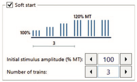
Soft start mode
Some treatment protocols guarantee an efficient stimulation at 110 or 120% of the MT. Such intensity may cause involuntary head movements in patients not familiar with this technique. To avoid such movements and prepare a patient, you can use the “soft start” included in the software. This allows you to start a stimulation at a low intensity and gradually and automatically increase it to the desired value.
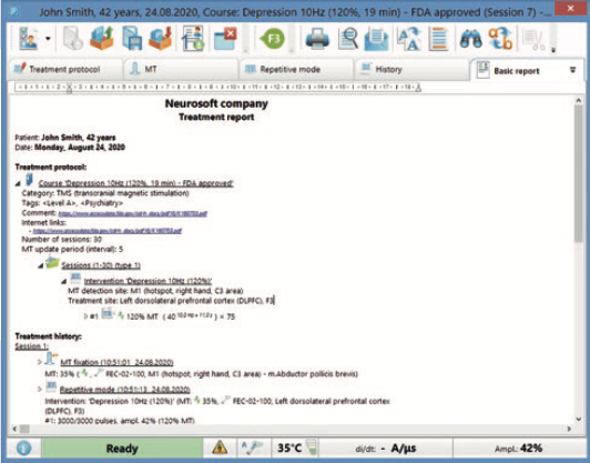
Report
Once the treatment session is completed, the software automatically generates a printable treatment report that includes the patient’s personal data, description of the treatment parameters and the entire treatment session history.